R&D
Spider silk protein scaffold
Use of spider silk protein-containing scaffolds as bone regeneration materials
Scaffold : Artificial ECM
Artificially made for transplantation and treatment of damaged tissues from diseases and injuries
Essential criteria of scaffold
-
Must be able to attach and deliver cells
-
Must induce cell proliferation and accelerate growth
-
Must stimulate cellular response
-
Must be quick and efficient with wound healing
-
Must be biocompatible and biodegradable, etc
Verification of Scaffold’s Properties
Market size
Biomaterial Market size
It is expected to reach USD 328.37 billion in 2027 from USD 109.39 billion in 2019
(CAGR 15.89%)
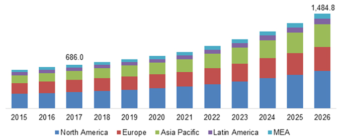
Scaffold Market size
The global scaffold technology market is valued at USD 728 million in 2018 and is expected to grow rapidly at a CAGR of 10.38%, reaching USD 1,485 million in 2026.
Scaffold technology market, By Region (2015 - 2026) (USD Million)
Spider silk
A biomaterial of high industrial interest due to
its excellent physical (strength, elasticity) and biological (biocompatibility, biodegradability) properties.
Mechanical properties
Balanced Strength & Extensibility
Functional textiles
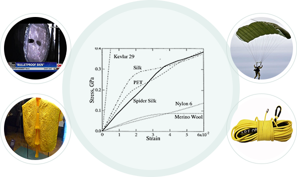
Stronger than Kevlar
for the same mass
Biological properties
Biodegradability & Biocompatibility
Biomedical applications
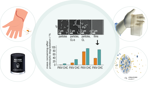
Excellent biocompatibility
& biodegradability (~6 months)
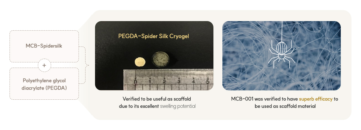
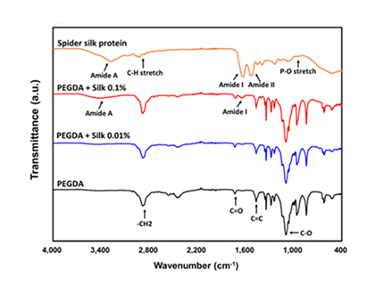
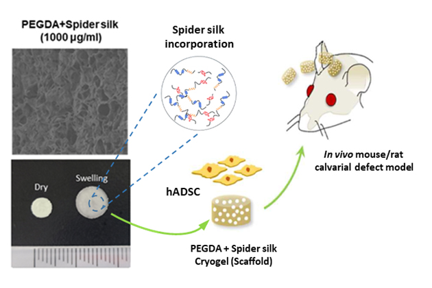
Spider silk protein-containing scaffold
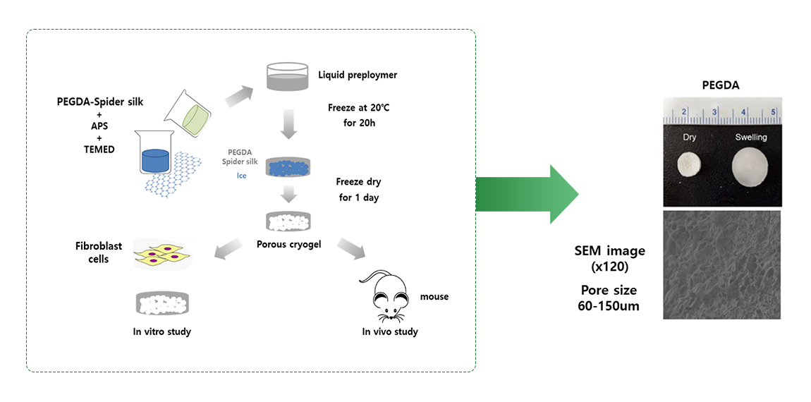
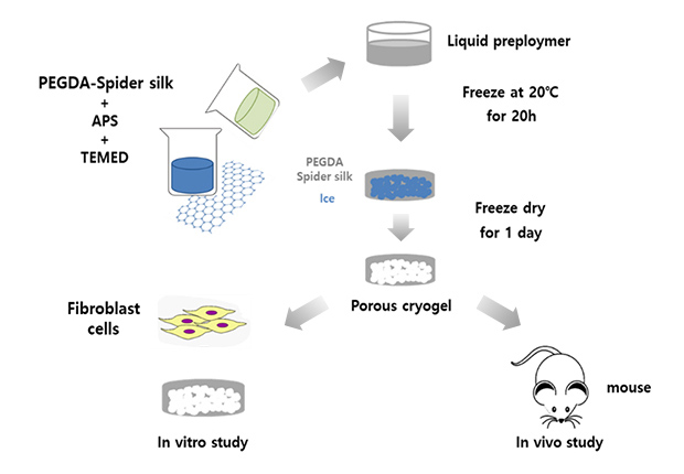
Development of a porous cryogel scaffold utilizing spider silk proteins.
By utilizing spider silk proteins possessing exceptional biological and physical characteristics, it is possible to address the shortcomings of existing metal and bio-scaffolds.
Furthermore, due to its porous structure, it excels in cell attachment and enables effective drug delivery, leading to enhanced cell growth and anticipated effects
in inducing stem cell differentiation. This can be applied to utilize it as a cell graft or bone graft.
Additionally, depending on the transplantation site, it can be freely shaped into various sizes and shapes.
Result
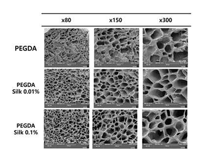
Fabrication of porous scaffold
Advantages of Porous Scaffolds
-
Easy cell attachment
-
Suitable for cell transplantation and induction of differentiation
-
Excellent growth of attached cells
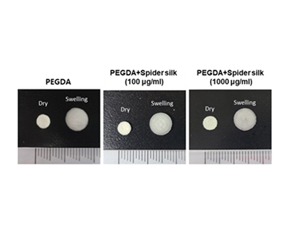
PEGDA Advantages
-
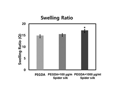
Excellent swelling ratio
-
Convenient for moisture retention, cell penetration, and drug delivery
-
Suitable material for inducing bone differentiation*
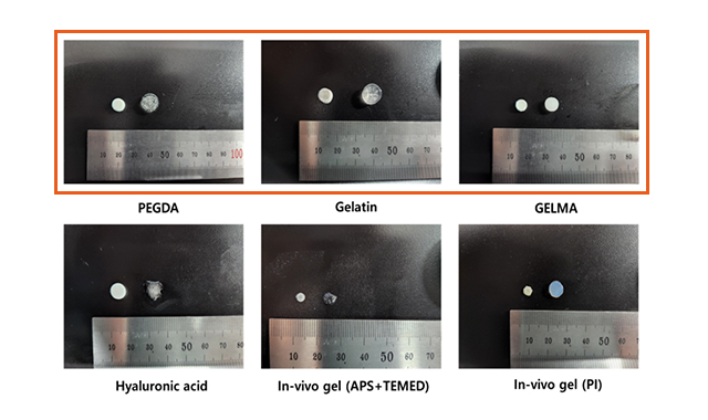
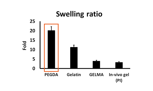
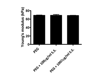
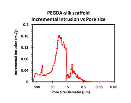
Mechanical properties of spider silk scaffold
Swelling ratio, compressive strength, porosity and pore size all show the possibility of spider silk protein to be used as scaffolds
Young’s modulus 64~68 kPa Porosity : >85% pore size : 20~200 um
(Adequate for cell attach & osteogenic differentiation*)
Live/Dead assay (Biocompatibility)
Biocompatibility of PEGDA-spider silk scaffold
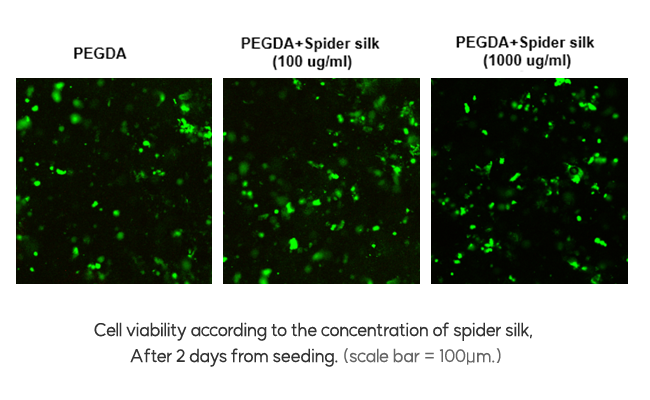
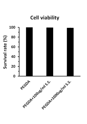
Confirmation of excellent biocompatibility of PEGDA-spider silk cryogel
-
After constant seeding of cells in the fabricated scaffold, live/dead assay was performed. (live cells show green fluorescence and dead cells show red fluorescence)
-
It was confirmed that the survival rate was more than 98% in scaffolds containing spider silk protein.`
PEGDA
spider silk scaffold cell adhesion

Cell adhesion according to the concentration of spider silk (Confocal image)
293T cells tagged with GFP were seeded on the scaffold at a constant concentration (2×10⁵ cells in a medium volume of 50 μL)
-
After 24h of seeding, as the scaffold swelled, it was checked whether the cells were properly attached and permeated evenly.
-
Cells are more and more evenly attached to PEGDA-cryogel containing spider silk protein. (Confocal imaging)
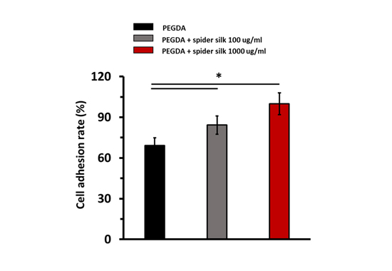
Cell adhesion rate on cryogel
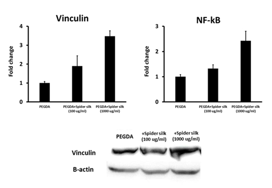
Expression of genes involved in focal adhesion and cell growth
In-vitro bone
regeneration efficacy
Calcium deposition
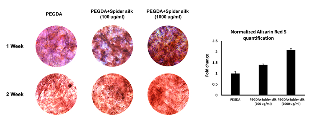
Alizarin Red S staining of hADSCs
Alizarin Red S staining showed higher calcium deposition in PEG+spider slik protein cryogel group when compared to control group
Osteogenic gene marker
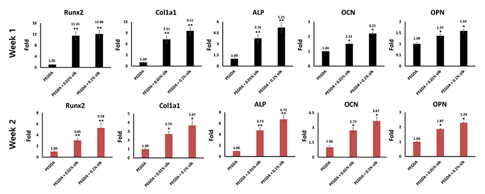
Osteogenic gene marker expresstion of hADSCs
Increase of osteogenic gene markers found in experimental group with spider silk protein
Bone regeneration improvement in vivo test
Rat calvarial defect model in vivo test
-
Transplantation of Spider silk incorporated scaffold into skull
-
Growth factor (BMP, Bone Morphogenetic Proteins) loading for bone regeneration
-
Confirm bone regeneration after 12 weeks (Micro CT, Histology, Immunohistochemistry etc.)
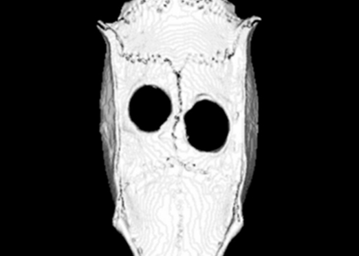
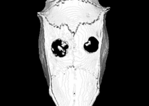
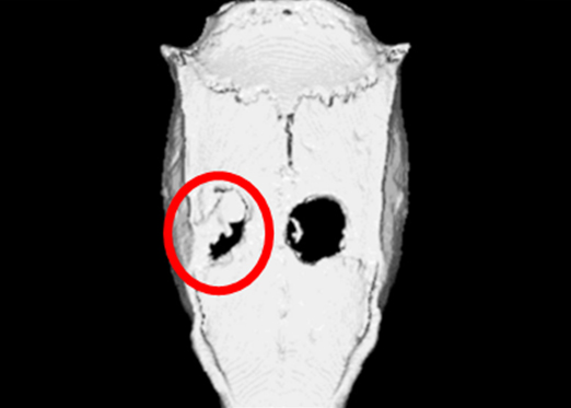
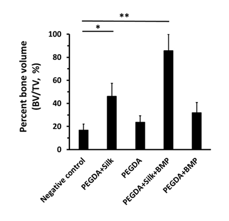
In-vivo bone
regeneration efficacy
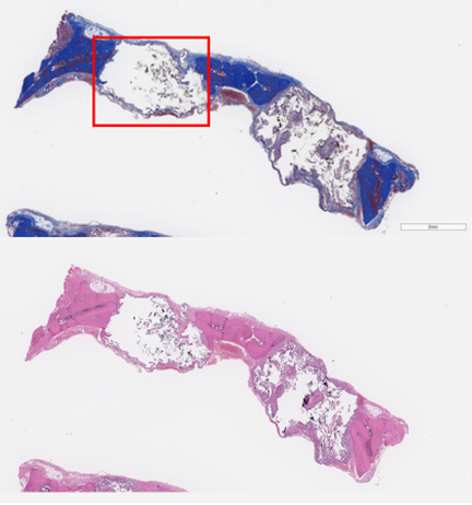
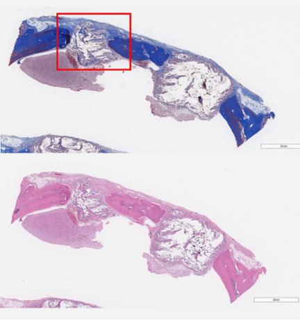
Rat calvarial defect model in vivo test
|
L Negative control R Negative control |
L PEGDA+Slik R PEGDA |
L PEGDA+Slik+BMP R PEGDA+BMP |
|---|---|---|
 |
 |
 |
 |
 |
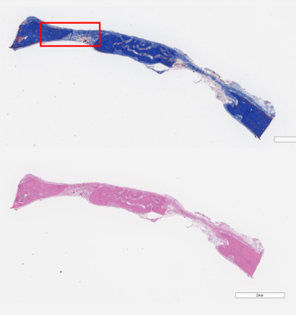 |
Histological analysis
Masson's trichrome staining
-
Red keratin and muscle fibers
-
Blue or green collagen and bone
-
Light red or pink cytoplasm
-
Dark brown to black cell nuclei
H&E(Hematoxy and eosin) staining
-
Hematoxylin stains cell nuclei a purplish blue
-
Eosin stains the extracellular matrix and cytoplasm pink
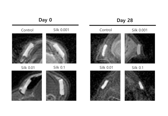
Confirmation of biocompatibility
and biodegradability
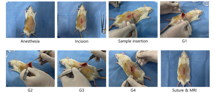
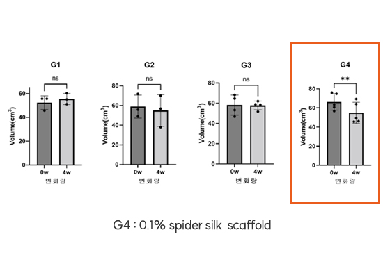
After 28 days of subcutaneous implantation, the extent of degradation and adhesion were assessed:
-
A significant enhancement in biodegradability was observed in the group containing 0.1% silk.
-
MRI imaging revealed a substantial reduction in volume of the implanted scaffold.
-
Tissue analysis including H&E staining confirmed biocompatibility of all test samples.